Cytotoxicity and biodegradation of cerium oxide-iron oxide nanoparticle platform as potential theranostic agent for ROS related diseases
Project Director: Dr. Valentin-Adrian MARALOIU
Accumulation of reactive oxygen species (ROS) causes the occurrence of numerous disorders, such as cancer, as well as cardiovascular, inflammatory and neurodegenerative diseases.
An efficient strategy against such diseases relies on efficient diagnostic of the incipient stages of development using imaging techniques, but also on effective therapeutic systems. Systems containing nanoparticles with combined diagnostic and therapeutic capabilities, called theranostic agents, represent a new strategy for reducing the economic burden for healthcare systems produced by the effects of these diseases.
This multidisciplinary project will determine the cytotoxicity and the biodegradation of cerium oxide-iron oxide nanoparticle platform. This will be done using an acid medium mimicking the biological environment, in vitro using various 2D and 3D culture systems and in vivo using a murine model. The follow-up of theranostic agents in biological media will be studied up to two weeks for the in vitro model and up to three months for the in vivo murine model using Transmission Electron Microscopy (TEM) techniques: conventional TEM and electron tomography for biolocalization and biodistribution of nanoparticles and their potential induced necrosis to cells; High Resolution TEM, Selected Area Electron Diffraction for biodegradation and biotransformation; Electron Energy Loss Spectroscopy combined with Scanning TEM for evolution of oxidation states of O, Fe and Ce.
The purpose of this multidisciplinary project is to evaluate the efficacy of the studied theranostic agents biodistributed in biological media using Transmission Electron Microscopy techniques.
The studied theranostic agents containing nanoparticles of iron oxide (maghemite - γ-Fe2O3) and cerium oxide will agglomerate, after their injection in mice, in macrophages inside phagolysosomes. The evolution of these nanoparticles will be followed on a long period of time up to 120 days after administration. In time, because of the acidic medium from phagolysosomes, the nanoparticles will be degraded, most of them will change their crystalline structure. This biotransformation will be evidenced by electron diffraction patterns and EELS spectra.
These results will be compared with the ones obtained in an acidic environment mimicking the biologic one, as well as in 2D and 3D cancerous cell cultures.
Dr. Valentin-Adrian Maraloiu - Principal Investigator
Dr. Raluca Florentina Negrea - Postdoc
Drd. Catalina Gabriela Mihalcea - PhD Student
Drd. Cristian Radu - PhD Student
Phase I: Characterization of CO-ION platform with controlled size and geometry and its biodegradation in an environment mimicking the biological one
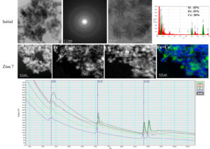
Starting from Fe3O4 and CeO2 nanoparticles with determined morpho-structural characteristics, several CO-ION platforms were synthetized in which the thickness of the CeO2 shell around Fe3O4 nanoparticles varied. Some of these platforms were thermally treated. Using Transmission Electron Microscopy techniques (CTEM, SAED, HRTEM EDS si EELS), morphology, crystalline structure and chemical composition of nanocomposites before and after thermal treatment were determined. It was proved that thermally untreated nanocomposites kept the initial characteristics of Fe3O4 and CeO2 nanoparticles. In the case of thermally treated nanocomposites, some changes of morphological characteristics of initial Fe3O4 and CeO2 nanoparticles were observed: Fe3O4 nanowires disappeared and the size of Fe3O4 and CeO2 nanoparticles slightly increased.
The degradation of the chosen CO-ION platform was studied using an acidic medium similar with the one found in lysosomes. The biodegradation experiments were conducted in citric acid at various values of pH and certain time periods after the insertion of nanocomposites in acidic medium. The investigations carried out using CTEM, SAED, HRTEM EDS and EELS techniques demonstrated the acidic medium didn't provoked changes of morphology, crystalline structure or chemical composition of nanocomposites in the period of 3 weeks.
Phase II. Biodegradation of CO-ION platform in vitro si in vivo
The second phase of the project took place between January 1 – December 31 2023, in the frame of 6 activities. This phase was dedicated to the determination of the way in which biodegradation of CO-ION platform take place in vitro and in vivo at certain time intervals after internalization.
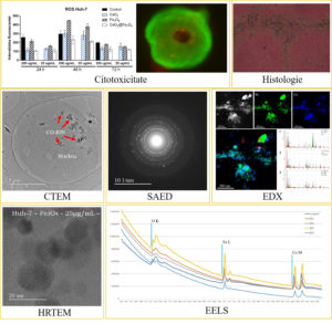
For the study of biodegradation of CO-ION platform in vitro 2D and 3D (spheroids) cell cultures were used starting from liver (Huh7) and kidney (Vero) cell lines. Cultures were incubated with initial nanoparticles of O4 and CeO2, as well as CO-ION platform, at two concentrations (25 µg/mL and 200 µg/mL) for 1, 2, 3 and 5 days. Both cytotoxicity analyses and the results obtain from Transmission Electron Microscopy techniques demonstrated the fact that at low concentrations studied nanoparticles are not toxic, while at high concentrations there is a small toxicity. TEM images showed that the distribution of nanoparticles in cells is heterogenous, that they are located only in cytoplasm once they are internalized by cells. TEM techniques also demonstrated that there are no crystallinity changes in 5 days (for 2D cultures) or in 15 day (for spheroids).
For the study of biodegradation of CO-ION platform in vivo C57BL/6 mice were used intravenously injected with CO-ION platform with a dose of 25 ug/mL. Ex vivo samples from different organs (liver, kidney, spleen, brain) at different time intervals (1, 7, 15, 30, 60, 90 and 120 days) after injection were prepared by fixation, postfixation, dehydration and resin imbedding, sections of 100 nm thickness from resin blocks being cut using Leica UC7 ultramicrotome.
TEM images showed that the distribution of nanoparticles in cells and organs is heterogenous, that they are located in cytoplasm. On the other hand it can be observed that in time the size of CO-ION platform agglomerations decreases and that the agglomeration become more dispersed. Also, ferritin agglomerations can be observed which it can be intrinsic (as a result of normal metabolic processes) or extrinsic (as a result of the biotransformation of Fe3O4 nanoparticles in ferritin). Also, in CTEM images no sign of toxicity induced by the presence of nanoparticles in organs can be observed.
Phase III. Dissemination of the results
Beside the article published in 2023, there are three other manuscripts there are being prepared:
- “Fe3O4/CeO2 Nanoplatform with “Core/Shell”-Like Structure: biodegradation and biotransformation in mimicking intracellular environment”; authors: Valentin-Adrian Maraloiu, Catalina Mihalcea, Cristian Radu, Yuliia Shlapa, Anatolii Belous;
- “Cytotoxicity and biodistribution of Fe3O4/CeO2 Nanoplatform with “Core/Shell”-Like Structure in 2D / 3D healthy cell cultures”; authors: Valentin-Adrian Maraloiu, Catalina Mihalcea, Cristian Radu, Aida Selaru, Sorina Dinescu, Yuliia Shlapa, Anatolii Belous;
- “Biodegradation and biotransformation of Fe3O4/CeO2 Nanoplatform with “Core/Shell”-Like Structure in murine model” authors: Valentin-Adrian Maraloiu, Catalina Mihalcea, Cristian Radu, Cosmin Mustaciosu, Yuliia Shlapa, Anatolii Belous.
The results of phase II will be presented in a poster presentation at the 9th International Workshop of Materials Physics which will take place between May 14 - May 16 2024 in Magurele, Romania. The presentation will be entitled „Cytotoxicity and biotransformation of Cerium Oxide-Iron Oxide platform in cells cultures and murine model”.
Published articles:
1. Belous, A - Design of Magnetic Fe3O4/CeO2 "Core/Shell"-Like Nanocomposites with Pronounced Antiamyloidogenic and Antioxidant Bioactivity; ACS APPLIED MATERIALS & INTERFACES; 15 (42); 49346-49361; DOI: 10.1021/acsami.3c10845
Conferences:
1. Oral presentation: "Biodistribution, biodegradation and cytotoxicity of Cerium Oxide-Iron Oxide platform in healthy cells",
Authors: Valentin-Adrian MARALOIU, Yuliia Shlapa, Anatolii Belous, Aida Selaru, Sorina Dinescu
5-th Conference of Romanian Electron Microscopy Society, 18-27 Octomber 2023, Cluj-Napoca, Romania
2. Poster presentation: "Cytotoxicity and biotransformation of Cerium Oxide-Iron Oxide platform in cells cultures and murine model"
Authors: Valentin-Adrian Maraloiu, Catalina Mihalcea, Cristian Radu, Yuliia Shlapa, Anatolii Belous, Aida Selaru, Sorina Dinescu, Cosmin Mustaciosu
9-th International Workshop of Materials Physics, 14-16 May 2024, Magurele, Romania
Valentin-Adrian Maraloiu
Researcher II
National Institute of Materials Physics
Laboratory of Atomic Structures and Defects in Advanced Materials
405A Atomistilor str., PO Box MG. 7
Magurele - Bucharest, Romania
077125
Telephone: +40212418241
Fax: +40213690177
PROJECTS/ NATIONAL PROJECTS
Copyright © 2026 National Institute of Materials Physics. All Rights Reserved